
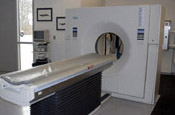

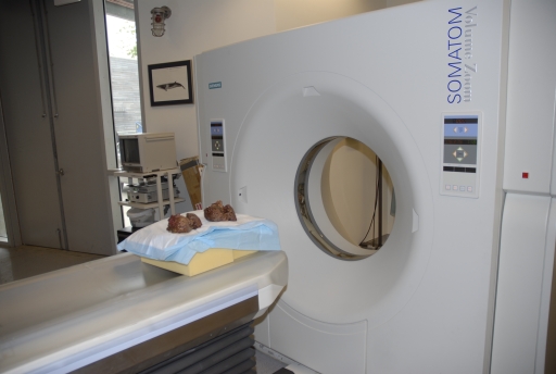
Siemens ® Somatom Volume Zoom CT Scanner Specifications
 |
 |
 |
 |
 |
 |
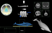
Spiral Scanning Technique: (See how it works)
- Ultra-fast scanning technique with continuous table feed
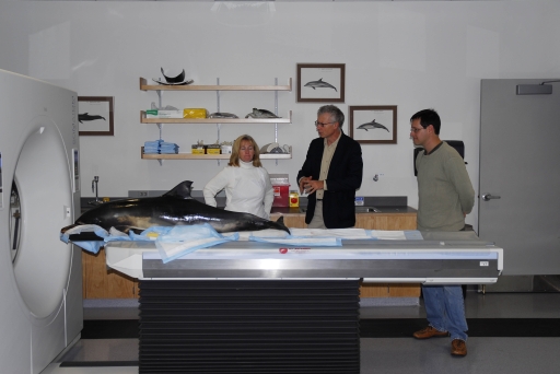
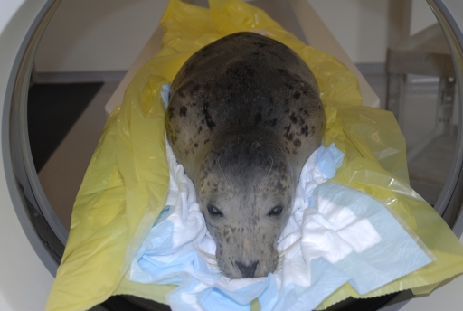
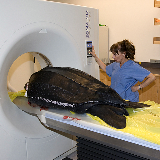
- Up to 80 mm/s volume coverage for minimized examination times for live animals
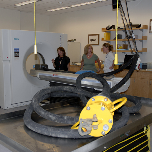
- Data acquisition of an entire anatomical volume up to 157 cm without pause
- Maximum Table Load 200 kg / 450 lbs
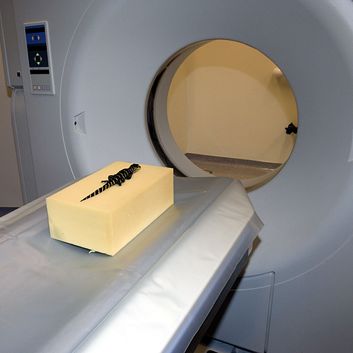
- Maximum Gantry Aperture 70 cm.
- Maximum FOV Diameter 55 cm
- Slice Thickness/Collimation 0.5 mm - 10 mm
- Slice Increments 0.1 mm - 10 mm (increment is the distance between the center of adjacent images)
- Scanner Table Pitch 1 - 8. (pitch is the ratio of table feed per rotation to collimation of a single slice)
- Topogram Data:
Min/Max Length: 128/1024 mm
Min/Max Time: 1.6/10.6 sec
Views: Anterior to Posterior (AP), Posterior to Anterior (PA), Lateral (LAT)
Sequence Scanning Technique: (related publication)
- Ultra fast acquisition with or without table feed
- Automatic clustering of scans
| Sequence Slice Width | ||
| Collimation (mm) | Thickness (mm) | |
| 2 x 0.5 | 0.5 | |
| 4 x 1.0 | 1.0 | |
| 4 x 2.5 | 2.5 | |
| 4 x 5.0 | 5.0 | |
| 2 x 8.0 | 8.0 | |
| Combi Scan | ||
| Collimation (mm) | No. slices fused | Thickness (mm) |
| 2 x 0.5 | 2 | 1.0 |
| 4 x 1.0 | 2 | 2.0 |
| 4 x 1.0 | 4 | 4.0 |
| 4 x 2.5 | 2 | 5.0 |
| 4 x 5.0 | 2 | 10.0 |
Image Reconstruction:
- 125 ms temporal resolution
- 30 lp/cm spatial resolution
- Scan fields 50 cm; 25 cm (ultra-high resolution collimator)
- Reconstruction Field 5 - 50 cm
- Reconstruction Time 1.5 images/sec
- Matrix 512 x 512 pixels
- Hounsfield Units Scale -1024 to +3071 HU (Extended Scale -10240 to + 40960 HU)
- Tube Current Range 28 - 500 mA
- Tube Voltage 80120, 140 kV
- DICOM Format Images
- Multiplanar Reformations (MPR)
- Maximum/Minimum Intensity Projections (MIP/MinIP)
- Shaded Surface 3D Reconstructions (SSD).
- Volume Rendering Technique (VRT)
Siemens® DICOM Conformance Statement
DICOM Conformance Specifications:
A Nontechnical Introduction to DICOM: (Horii, 1997)
"The initial goal in developing a standard for the transmission of digital images was to enable users to retrieve images and associated information from digital imaging equipment in a standard format that would be the same across multiple manufacturers. The first result was the American College of Radiology (ACR)-National Electrical Manufacturers Association (NEMA) standard, which specified a point-to-point connection. However, the rapid evolution of computer networking and of picture archiving and communication systems meant that this point-to-point standard would be of limited use. Consequently, a major effort was undertaken to redesign the ACR-NEMA standard by taking into account existing standards for networks and current concepts in the handling of information on such networks. The Digital Imaging and Communications in Medicine (DICOM) standard was the result of this effort. Its popularity has made discussion, if not implementation, of the standard common whenever digital imaging systems are specified or purchased."
